


来自日本理化学研究所(RIKEN)与中国科学院西安光学精密机械研究所的联合研究团队,在极紫外(XUV)波段实现了孤立阿秒脉冲的亚微米级聚焦。研究团队采用一块定制的椭球面多层膜反射镜,将峰值功率为1.1-GW的桌面型孤立阿秒脉冲聚焦至0.46×0.36 μm大小(FWHM),聚焦后峰值强度达到3×10¹⁵ W/cm²。该成果为在气体与固体中开展阿秒非线性光学研究奠定了实用化基础。
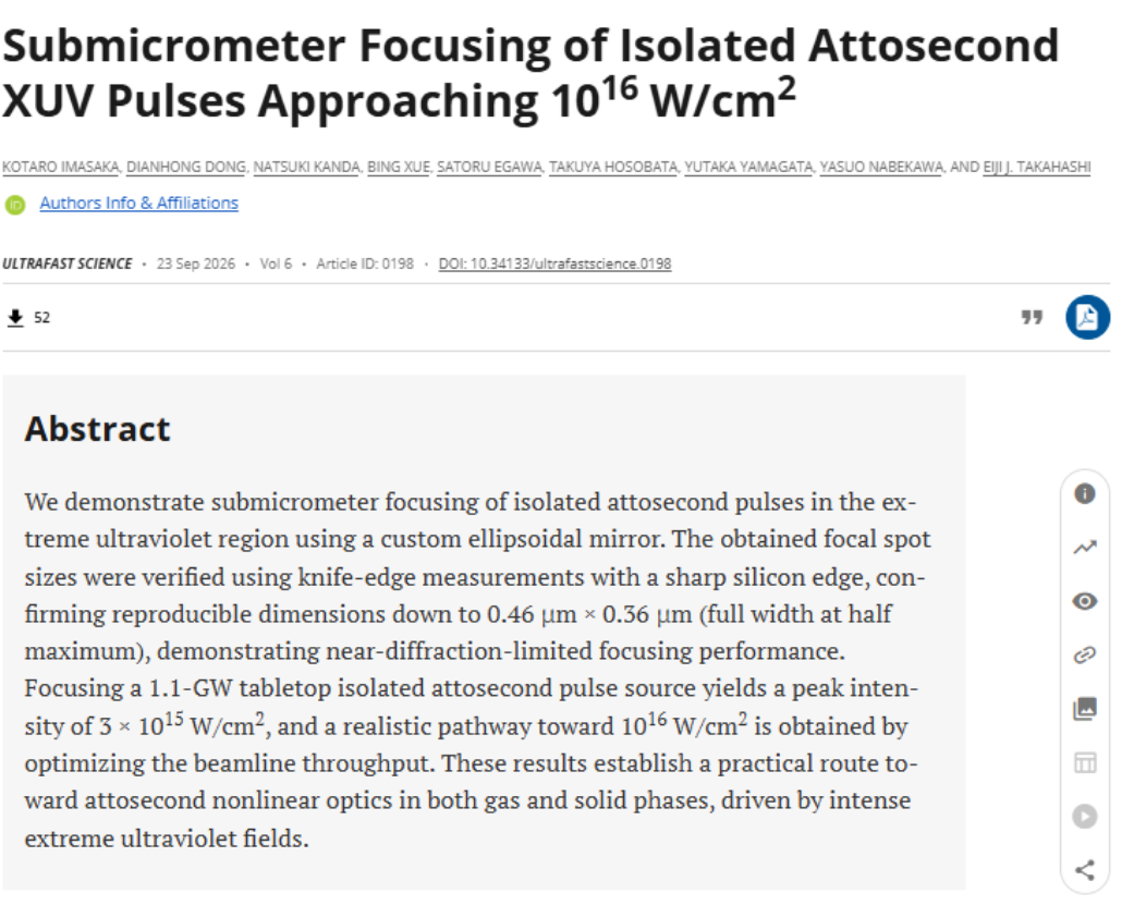
阿秒(as)是十亿分之一秒的十亿分之一(即10-18秒),阿秒激光脉冲的持续时间通常为几十至几百阿秒,为在电子运动的自然时间尺度上直接观测电子行为提供了重要手段。若要利用阿秒脉冲进一步操控电子运动,甚至直接驱动非线性光–物质相互作用,焦点处还需要足够高的峰值强度;在脉冲能量和脉冲宽度一定时,把脉冲聚焦到更小的焦斑,是提高峰值强度最直接的途径。此前,研究人员已实现阿秒脉冲串(由一系列重复的阿秒脉冲组成)的亚微米聚焦;而孤立阿秒脉冲仅包含一个波形明确的脉冲,因此更适用于阿秒泵浦-探测等超快时间分辨实验——但要将这样的脉冲聚焦到足够小的焦斑、获得足够高的强度,长期以来一直面临较大的技术挑战。
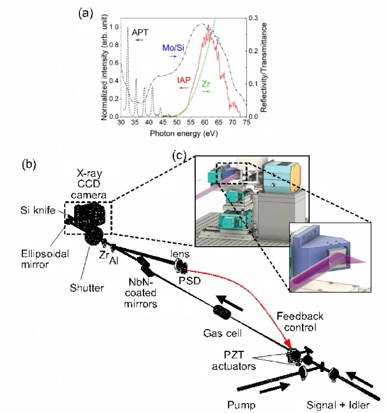
Fig. 1. Spectral conditions and experimental setup for XUV focusing. (a) Spectralconditions relevant to the focusing experiments. The APT (black dashed line) and IAP(red solid line) spectra at the XUV focus are shown, along with the transmission of a 100-nm-thick Zr thin-film filter (green dotted line) and the reflectivity of the Mo/Si multilayer coating for s-polarized light (blue dashed–dotted line). The Zr-filter transmission was calculated using the optical constants reported in Refs. [34,35]. (b) Schematic of HHG beamline. The system comprises waveform synthesis, HHG in a gas cell, and XUV beam transport. (c) Enlarged view of the focusing region. The incident beam is drawn thicker than the scale for clarity.
为攻克这一难题,联合研究团队开发了一套基于椭球面多层膜反射镜的聚焦系统,该反射镜专门针对中心光子能量约60 eV的XUV光设计。在利用刀口法测量聚焦光斑的过程中,研究团队采用位置反馈装置稳定了XUV焦点位置,并引入基于中位数的统计分析方法,抑制残余焦点位置波动带来的测量误差,从而在真实实验条件下实现了对亚微米焦斑尺寸的可靠定量评估。
实验测得的最小焦斑尺寸为水平方向0.46±0.13 μm、竖直方向0.36±0.12 μm,接近波动光学模拟所预测的衍射极限性能。对于峰值功率1.1-GW的孤立阿秒脉冲光源而言,在考虑束线的光学损耗后,这一焦斑尺寸对应的峰值强度达3.0×10¹⁵ W/cm2。据研究团队所知,这是首次在实验上实现基于高次谐波产生(HHG)的孤立阿秒脉冲的亚微米聚焦,且光斑尺寸测量经过定量验证;其3.0×1015 W/cm2的峰值强度,也是迄今已报道的桌面型HHG孤立阿秒脉冲光源中的最高纪录。
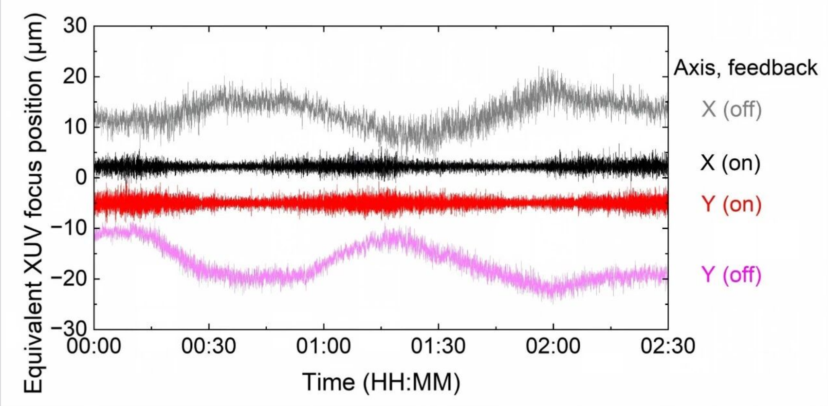
Fig. 2. Performance of focal-position stabilization system. Temporal traces of the PSD-derived focal-position signal in the horizontal (X) and vertical (Y) directions with and without feedback.
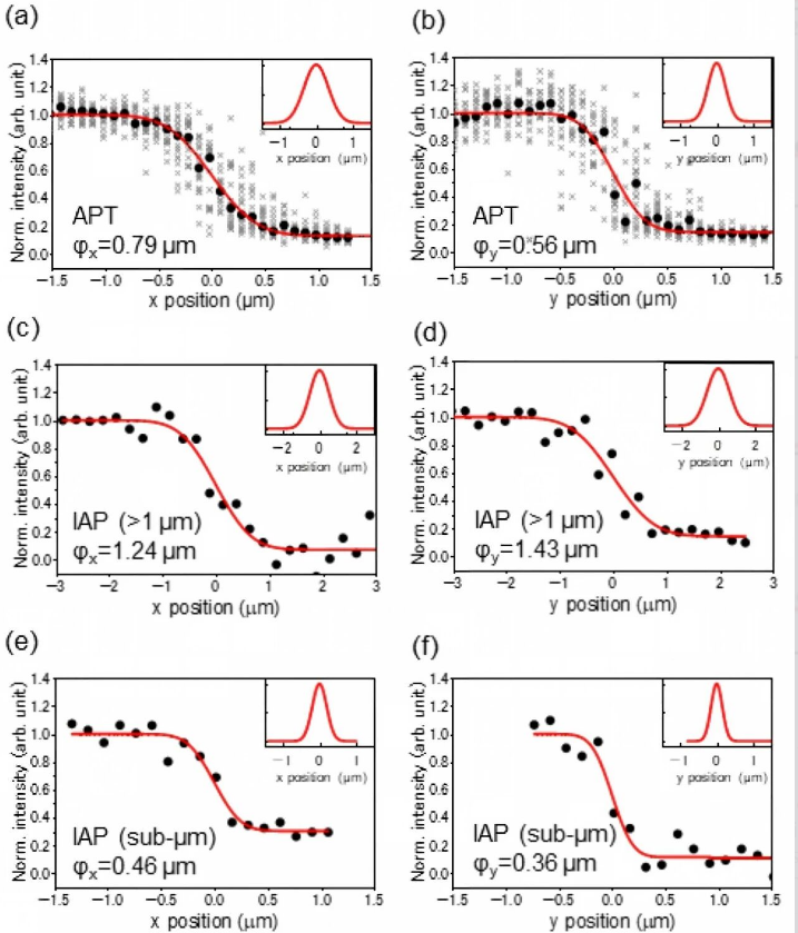
Fig. 3. Knife-edge characterization of focused XUV beam. (a) and (b) APT measurements in the horizontal (X) and vertical (Y) directions, respectively. (c) and (d) IAP measurements showing spot sizes slightly above 1 μm. (e) and (f) IAP measurements demonstrating submicrometer focusing. The gray cross markers represent raw accumulated signals, the black circles represent the median-processed intensity values, and the red lines represent the error function fits. The insets show the corresponding reconstructed beam profiles obtained from the derivative of the fitted error functions, representing Gaussian beam profiles.
这一成果为阿秒非线性光学在气态、固态及其他凝聚态体系的拓展,提供了一条富有前景的研究路径。通过进一步降低阿秒束线中的光学损耗,该研究还指明了一条切实可行的路线,有望将聚焦强度提升至约2.6×10¹⁶ W/cm2,为进一步开展全XUV阿秒泵浦-探测实验奠定基础——将高峰值强度与阿秒级时间分辨能力相结合,深入探究超快电子动力学过程。
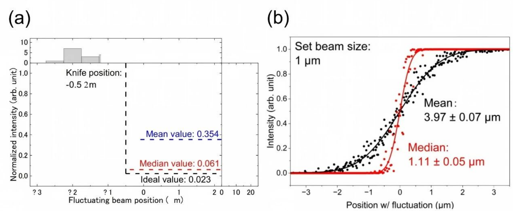
Fig. 4. Influence of statistical estimators on knife-edge beam reconstruction under residual focal fluctuations. (a) Simulated transmitted intensities as a function of beam position at a representative knife position, along with the corresponding histograms of beam-position fluctuations (top) and transmitted intensities (right). (b) Reconstructed beam profiles using mean and median estimators. Markers indicate the reconstructed data points, and solid lines represent error function fits.
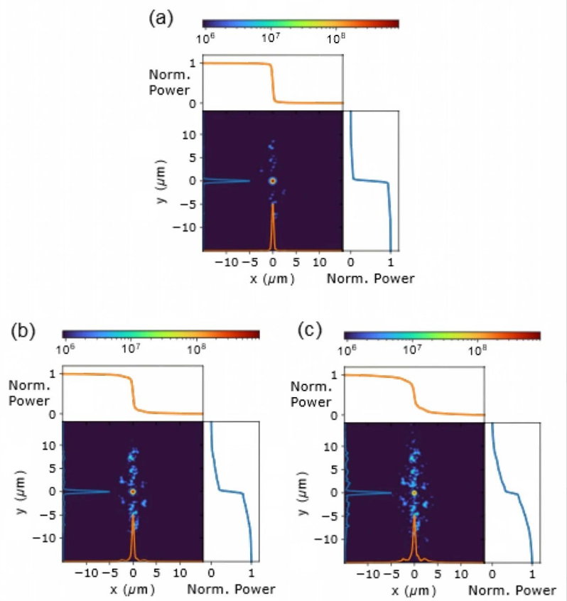
Fig. 5. Wave-optical simulations of the influence of local mirror surface variations on the focal spot size. (a) Measured surface map (1× roughness). (b) Surface map with twofold surface height variations. (c) Surface map with threefold surface height variations. For each case, the central panel shows the calculated 2D focal intensity distribution (logarithmic scale). Projection profiles (integrated along the orthogonal direction) are shown within the 2D maps (bottom and left axes). The upper and right panels show the corresponding knife-edge profiles used for beam-size reconstruction.
该研究由日本理化学研究所光量子工学研究中心(RIKEN, RAP)与中国科学院西安光机所超快光科学与技术全国重点实验室超快激光产生与调控研究中心组成的联合研究团队(XIOPM–RAP联合研究室)完成。Kotaro Imasaka、Dianhong Dong(董殿宏,西安光机所)和Natsuki Kanda为共同第一作者,并与其他合作者共同开展了实验和数据分析。中国科学院西安光机所薛冰研究员与日本理化学研究所极端光子学研究团队负责人Eiji J. Takahashi共同提出了实验构想与设计,实验工作主要在RIKEN完成,并由Eiji J. Takahashi负责项目指导。

 陕公网安备 61019002001027号