Optical microscopy is an indispensable tool for modern biomedical research. However, optical microscopic imaging of thick biological samples has always been a challenge. On the one hand, the resolution of microscopy systems is limited by the optical diffraction limit, which can only reach a maximum of about 200nm and cannot resolve sub-cellular details; on the other hand, biological samples are generally relatively thick and often exceed the depth of field of the microscope objective, and even in-focus targets within the depth of field are severely disturbed by out-of-focus light, resulting in very poor imaging signal-to-noise ratio. To address this problem, Yao Baoli's group at the Xi'an Institute of Optical Precision Mechanics (XIOPM) of the Chinese Academy of Sciences(CAS) has proposed an optical super-resolution and optical sectioning fusion technique for structured light-illuminated visible micro-imaging (iSIM). iSIM combines the existing super-resolution SIM (SR-SIM) and optical sectioning SIM (OS-SIM) techniques in a theoretical and methodological fusion, breaking the diffraction limit to achieve super-resolution imaging. The iSIM combines the theoretical and methodological fusion of two existing techniques, SR-SIM and OS-SIM, to break the diffraction limit and achieve super-resolution imaging, while obtaining a high signal-to-noise ratio optical slice image that reconstructs the 3D surface topography of the sample.
The present work starts from a theoretical model of SR-SIM imaging, to which an out-of-focus background interference term is added. Through detailed derivation, the role of the out-of-focus background interference term in the SR-SIM spectrum expansion is revealed - it only affects the intermediate spectrum at low frequencies, but not the extended high frequency spectrum (Figure 1); and the derivation also reveals the existence of equivalent mathematical expressions for the extended high frequency spectrum and OS-SIM images, thus establishing a theoretical link between SR-SIM and OS-SIM. The theoretical link allowing the imaging advantages of the two techniques to be fused (iSIM), resulting in super-resolved optical slices.
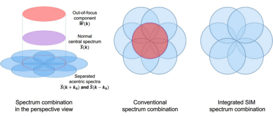 Figure 1. iSIM spectrum fusion method vs. conventional method schematic
In this work, a microscopic imaging simulation model was designed to verify the validity of the iSIM theory. The model starts at the vertical midline of the Ground Truth image and gradually blurs towards the ends of the image, with the degree of blurring proportional to the distance away from the midline. Based on this model, the simulation produces in-focus and out-of-focus effects due to the depth of field of the objective lens. By implementing iSIM, normal wide-field, OS-SIM and SR-SIM imaging on the model, the comparison demonstrates the imaging benefits of iSIM in terms of both super-resolution and avoidance of out-of-focus interference (Figure 2).
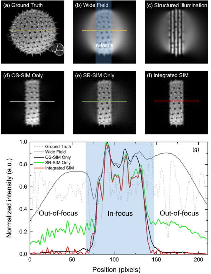 Figure 2. iSIM versus simulated imaging with normal imaging, OS-SIM, SR-SIM
The experiments further validate the imaging validity of the iSIM theory. The experimental optical path uses a spatial light modulator (SLM) loaded with grating phase to interfere with the diffracted ±1 level beam to produce structural illumination stripes.
To achieve the required stripe direction rotation for SIM and to simplify beam polarisation state control, the beam was experimentally modulated to interfere in a circularly polarised state (Figure 3). Using human retinal pigment epithelium (HRPE) as the sample, iSIM imaging was confirmed to combine the super-resolution advantages of SR-SIM with the high signal-to-noise ratio light sectioning capability of OS-SIM by comparison with normal wide-field, OS-SIM and SR-SIM imaging (Figure 4).
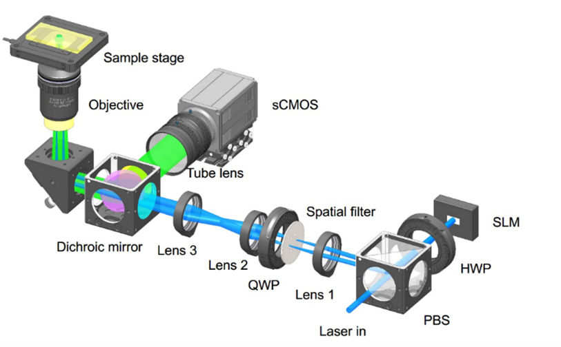 Figure 3. Experimental optical path diagram
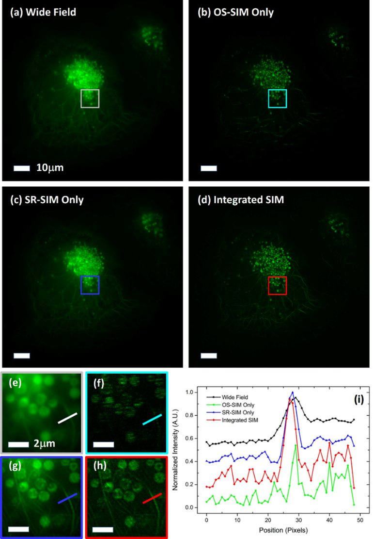 Figure 4. iSIM versus normal imaging, OS-SIM and SR-SIM imaging
 陕公网安备 61019002001027号