Fast high-resolution 3D imaging is important to achieve a complete and accurate understanding of biological processes due to the 3D nature of biological structures. Laser scanning confocal microscopy, namely the sequential illumination of restricted regions of specimens and rejection of the out of focus background with spatial filters has become an essential optical section and 3D imaging technique in contemporary biomedical research.
The super-resolution radial fluctuations (SRRF) approach has shown great promise in obtaining images with an improved spatial resolution by generating radiality maps from time-lapse frames. Unfortunately, in practice, SRRF often requires about 100 consecutive excitations to calculate the final result, which in turn leads to an increase of imaging time due to the large amounts of 3D imaging data acquisition, while suffering from an increased risk of photo-bleaching and photo-toxicity.
A research team led by Prof. YAO Baoli from the Xi'an Institute of Optics and Precision Mechanics of the Chinese Academy of Sciences proposed a novel deep learning network for 3D imaging of spinning-disk confocal microscopy (SDCM), named the SRRF-Deep. This network builds a 2.5D model with context information across neighboring pixels in z-scanning to facilitate the reconstruction of an optical slice in 3D imaging.
The SDCM imaging experiments were implemented using a commercial microscope. SRRF-Deep was designed to formulate an inverse image formation model by means of training with a data pair consisting of an SDCM stack and the corresponding SRRF imaging data.
Rather than treating the whole image as input, imaging frames are the first split and shuffled into a series of center-to-center sub-image pairs to avoid large training and computational costs.
Finally, the raw SDCM image stack was input into the well-trained model, which subsequently predicted a corresponding high-quality image. The results were published in IEEE PHOTONICS TECHNOLOGY LETTERS.
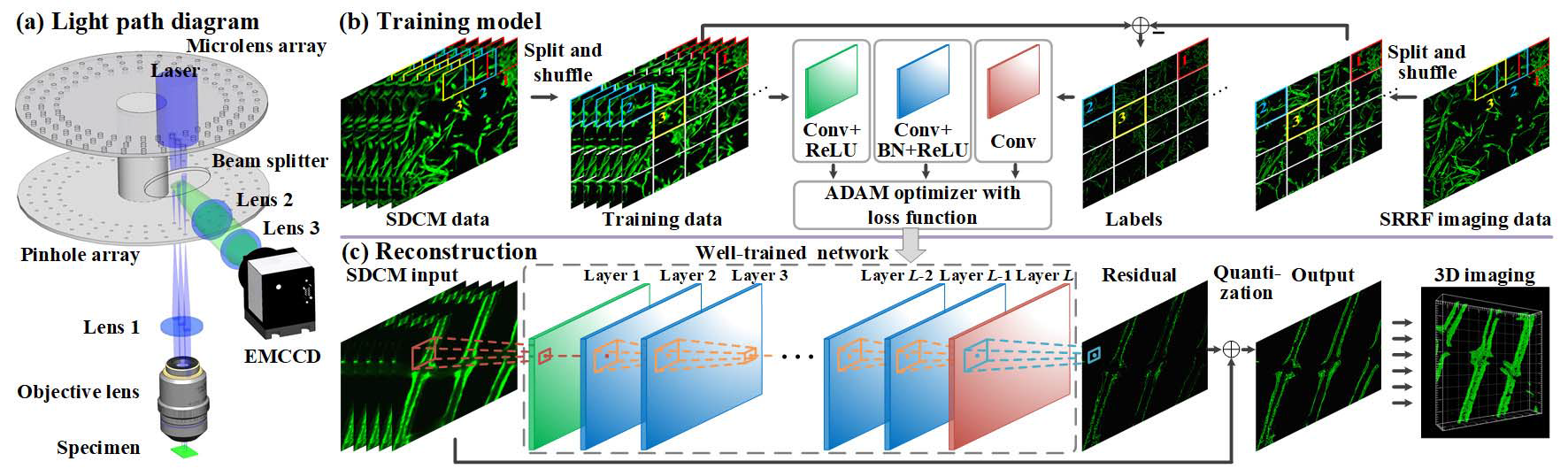
The proposed image system, training and reconstruction method. (Image by XIOPM)
By utilizing the proposed method, the researchers performed the experimental comparison between SDCM, SRRF and SRRF-deep. The experiments on daisy pollen, aspergillus conidiophores, onion pollen and mouse intestine showed high effectiveness and efficiency.
Compared to SRRF, SRRF-Deep reduces the number of scans required by a factor of 100 while accelerating the reconstruction of each slice by about 30 times.
Overall, this technique significantly improves the imaging throughput of the SRRF system by reducing both scanning requirements and reconstruction time, which makes the 3D imaging restoration faster and clearer.
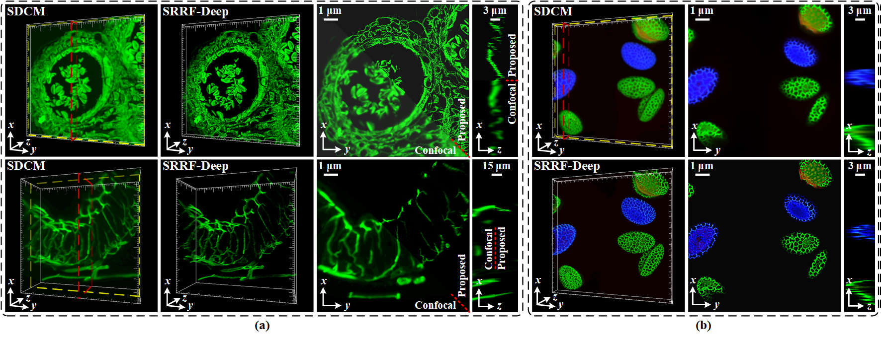
Real imaging examples with specimens obtained using the SDCM and SRRF-Deep methods. (Image by XIOPM)
Editor: ZHANG Nannan | Nov 04, 2020
 陕公网安备 61019002001027号